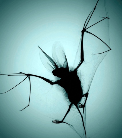
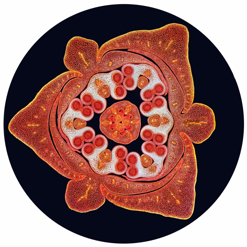
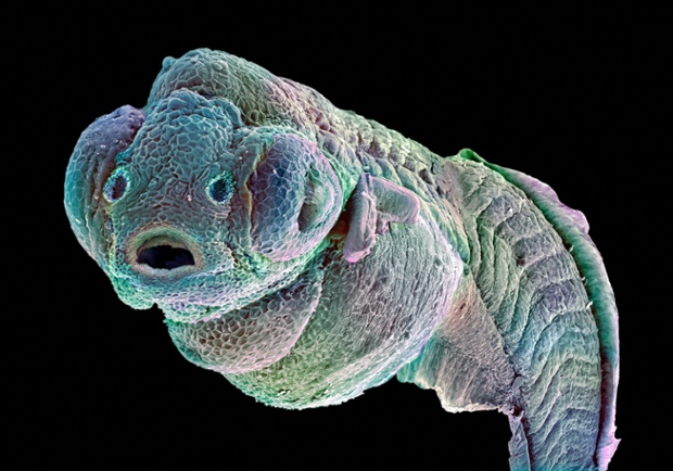
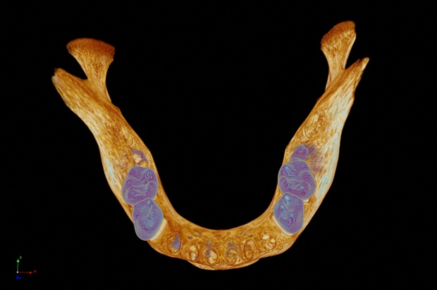
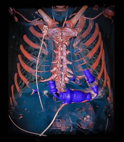
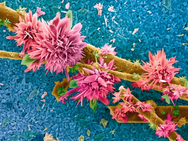
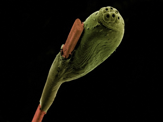
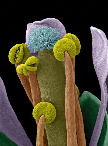
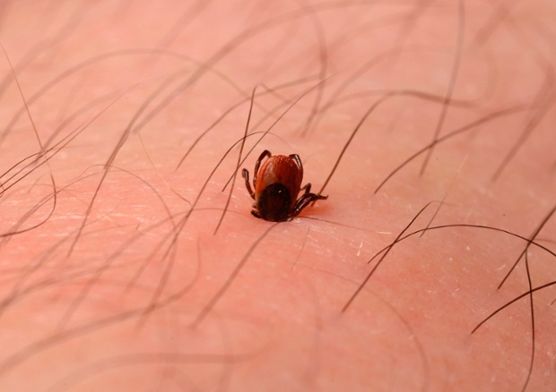
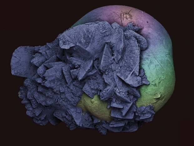
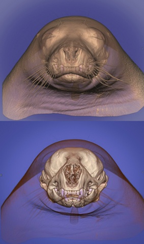
X-ray of a brown long-eared bat. Photograph: Chris ThornPhotograph: Chris Thorn/Wellcome TrustTransverse section through a stained lily bud showing the male and female reproductive organs. Photograph: Spike WalkerPhotograph: Spike Walker/Wellcome ImagesFalse-coloured scanning electron micrograph of a zebrafish embryo. Photograph: Annie Cavanagh & David McCarthyPhotograph: Annie Cavanagh/Wellcome ImagesBird's eye view of a model of a medieval human mandible, captured using a micro CT scanner. Photograph: Kevin Mackenzie/University of AberdeenPhotograph: Kevin McKenzie/University of Aberdeen/Welcome ImagesMechanical heart pump, as revealed by a dual energy tomography angiography of a human chest. Photograph: Anders PerssonPhotograph: Anders Persson/Wellcome ImagesFalse-colour micrograph of an agricultural sludge sample after burning in an oxygen atmosphere. Photograph: Eberhardt Josue Friedrich Kernahan & Enrique Rodriguez CanasPhotograph: Eberhardt Josua Friedrich Kernahan/Wellcome ImagesFalse-colour scanning electron micrograph (SEM) of a head louse egg attached to a strand of human hair. Photograph: Kevin Mackenzie/University of AberdeenPhotograph: Kevin McKenzie/University of Aberdeen/Wellcome ImagesSEM of an Arabidopsis thaliana flower, commonly known as thale cress. Photograph: Stefan EberhardPhotograph: Stefan Eberhard/Wellcome ImagesPhotograph of a deer tick embedded in a man’s skin. Photograph: Ashley Prytherch/Royal Surrey County Hospital NHS Foundation Trust/Wellcome ImagesPhotograph: Ashley Prytherch/Royal Surrey County Hospital NHS Foundation Trust/Wellcome ImagesSEM of a kidney stone (nephrolithiasis) – 2mm wide. Photograph: Kevin Mackenzie/University of AberdeenPhotograph: Kevin McKenzie/University of Aberdeen/Wellcome ImagesComputed tomography scan of the head of a seal. The image was created with a 3D volume rendering technique in which the skeleton has been made opaque and the soft tissues semi-translucent to reveal the skull. Photograph: Anders PerssonPhotograph: Anders Persson/Wellcome ImagesPhotograph of Astrantia major, Hadspen Blood. Photograph: Dr Henry OakeleyPhotograph: Dr Henry Oakley/Wellcome Images